Fighting resistance to antibiotics
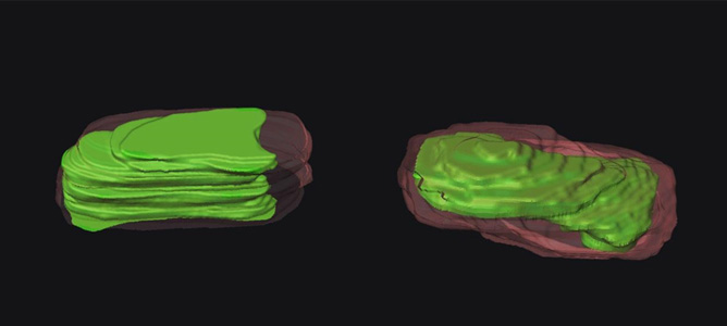
Reconstructed 3D models of single, untreated bacteria (left) and treated with 2 mg/L polymyxin B (right).
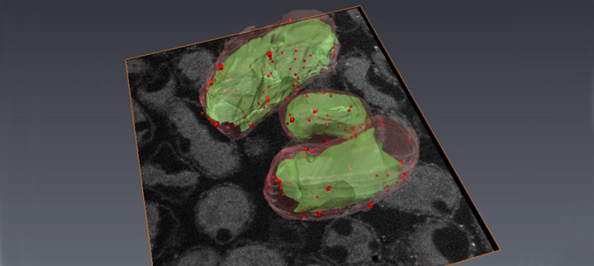
Three bacterial cells reconstructed using FIB-SEM tomography.
November 2014
As resistance to antibiotics becomes increasingly common, it is more important than ever to understand the mechanism by which antibiotics work on bacterial cells, and to develop new antibiotics which can be used against ‘super bugs.’
In order to understand the process by which potential combinations of antibiotics kill multi-drug resistant bacterial cells, a joint force from Monash Engineering (Mr. Boyin Liu and Dr. Jing Fu) and Monash Pharmaceutical Sciences (Professor Jian Li and Dr. Tony Velkov) together with the MCN, the Australian Synchrotron and the University of Queensland have been working on novel imaging approaches to assess the cellular responses of bacterial cells to the treatment of antibiotics, including the last-resort polymyxins.
The team has used the Focused Ion Beam (FIB) tool at MCN to mill away 25nm slices of a cell of resistant bacterial isolate (Klebsiella Pneumoniae) recently discovered in Queensland. After the removal of each slice, high-resolution scanning electron microscope images were taken and then reconstructed into a 3D model of a whole bacterial cell to reveal the effect of the antibiotic in different cellular regions.
The 3D models of both treated and untreated cells were reconstructed and compared. Their finding confirmed the invasion of polymyxin B on the cell envelope and the subsequent depletion of cytoplasmic materials. The results provided clear evidence for using rational antibiotic combinations to combat bacterial ‘superbugs.’
In the ongoing research, FIB is also being used to slice the bacterial cell to expose the interior surface, after which Atomic Force Microscopy (AFM) is employed to probe the intracellular changes and measure their mechanical properties. The investigators are also employing a single-molecule AFM tip functionalisation technique, Synchrotron imaging and secondary ion mass spectrometry to identify the chemical signatures due to antibiotic treatment. Their research is funded by the Australian NHMRC and the US National Institutes of Health (NIH).
You can read more about this project in In situ probing the interior of single bacterial cells at nanometer scale, published in IOP Science in September this year.

