High resolution x-ray imaging possible for delicate biological samples

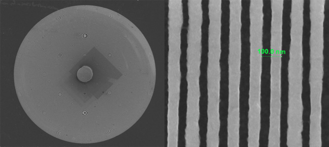
Top view of the 50nm resolution zone plate with the 30µm cross section. 

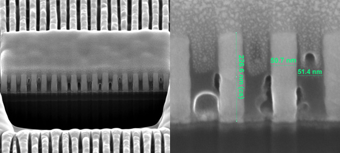
52o tilted view of the FIB cross-cut of the outer zones

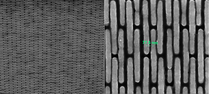
Top view of the 38nm resolution zone plate 

April 2014

High-resolution x-ray imaging is required to investigate processes in-situ or in-vivo samples or when it is particularly important that the sample is not damaged. This is possible due to the high penetration depth of the x-rays. Fresnel zone plates are optical elements that are capable of focusing x-rays with high efficiencies, allowing for nanometer size beams to deeply penetrate the imaging subjects.

A collaborative project between the MCN, La Trobe University and the Australian Synchrotron aims to fabricate high-resolution Fresnel zone plates and test their performance using synchrotron light.

As a starting point, a theoretical model was used in order to predict the highest achievable performance of zone plates for a given set of parameters. Subsequently, a fabrication protocol was chosen to produce these optical elements. The protocol involved testing a wide range of techniques including silicon wafer processing, photolithography, reactive ion etching, wet etching, electron-beam evaporation, electron-beam lithography, electro-deposition, and focused ion beam. The successful prototypes were tested at the Australian Synchrotron and their performance was measured against the theoretical predictions.

By utilising state-of-the-art instrumentation at the MCN to test theoretical predictions, the researchers have been able to create zone plates which exhibit properties that are close to the theoretical calculations. These zone plates are able to focus large diameter x-ray beams into nanometer-size spots which have high brilliance. This is important as it delivers not only high-resolution imaging capability, but it also minimises the exposure times which in turn maximises the data output capacity.

Furthermore, current prototypes incorporate in-built elements that act as x-ray beam stops, eliminating the need for additional stages usually required to mount external beam stops. This also limits the errors associated with the alignment process.

The benefits of using such optical elements are enormous as this is currently the only way to focus x-rays to nanometer-size beams with high efficiencies. This is paramount when high-resolution x-ray imaging is required to nondestructively investigate a diverse range of scientific problems.

The next stage of this project will be to produce prototypes on demand for the Australian Synchrotron scientific community to benefit diverse range of X-ray imaging techniques.