Nano-flowers for biosensing

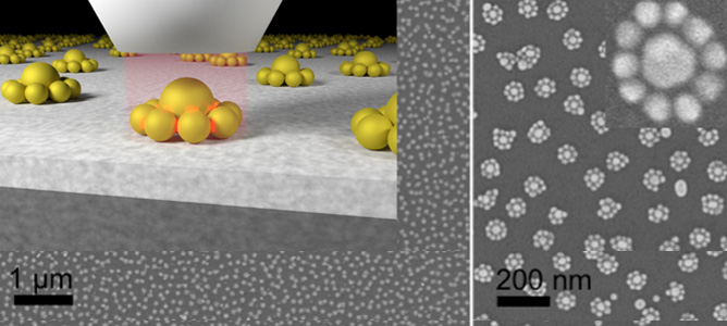
Schematic representation of the nano flowers focusing incoming light in the gaps. Overview SEM image on the left and zoomed-in views of the structures on the right showing the nano flower arrays. Credits: Soon Hock Ng, Yuanhui Zheng.

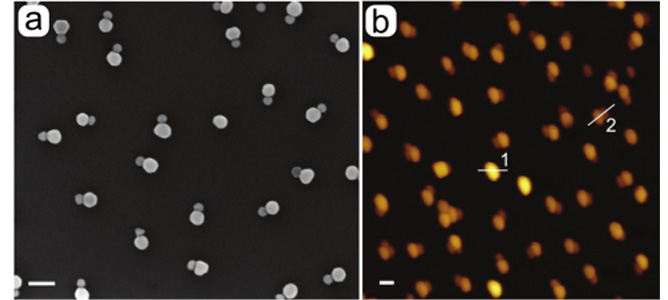
Pairwise assembled nanoparticles imaged with SEM (a) and AFM (b).

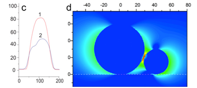
Vertically stacked particles can be found in the AFM images by using line scans in the AFM images (c). The yield of dimers with the here developed protocol is as high as 85%. (d) shows a calculation of the field focusing in the gap of such a dimer (red indicates a high intensity).

March 2015

Gold nanostructures separated by a small gaps (1/10,000 the thickness of a human hair) show a strong focusing effect of the electric field into this gap when exposed to light. Assembly of nanoparticles in flower structures with multiple of such gaps, have been developed. Using a spectroscopic technique known as Surface Enhanced Raman spectroscopy (SERS), these structures have the potential to detect minute quantities of molecules used in explosives or cancer markers in blood.

A team of researchers led by MCN Technology Fellow, Associate Professor Udo Bach from CSIRO have attached DNA onto gold nanoparticles to give them a negative charge. Small glass surfaces were given a positive charge, after which the glass is placed into the gold particle solution. This enables a regular distribution of particles on the surface. DNA requires two different parts to form a double helix - the DNA on the surface particles contains one such part, while a solution of smaller particles has the other part of the DNA attached to it. When the glass is placed into the solution with the smaller particles, the DNA comes together to form a double helix and connects the smaller particles to the larger ones, leading to the flower structure seen in the image above.

The assembly of these flowers was carried out in the MCN biochemistry laboratory, while the progress and results have been monitored using MCN’s UV-Vis, SEM and AFM tools.

In comparison to commercially available SERS sensor chips, the flower structures has proved to be superior in signal intensity and reproducibility. This is particularly interesting, as a new and simpler fabrication method for these complex structures has been used in this study.

Besides the improvements in molecular sensing, the techniques developed in this project may be used to control the assembly of nanoparticles of various materials and therefore applications in catalysis are envisaged, for example. This is particularly attractive in times where renewable energies and processes like water splitting are of high interest.

Future stages of the project aim to continue in the same vein, as electrostatic forces can be used to assemble nanoparticles in pairs. Similar hotspots as in the flower structures can be produced in an even simpler way, while the effect of the size and the material of the nanoparticles on the performance may help to unlock new applications for such structures.