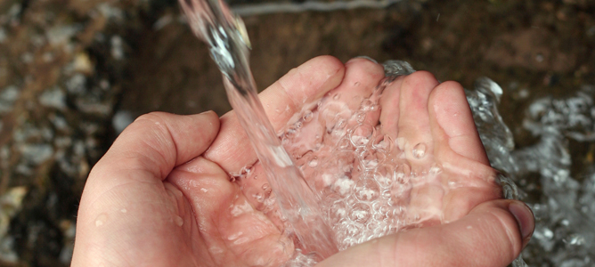
Protecting the nations’ water supply
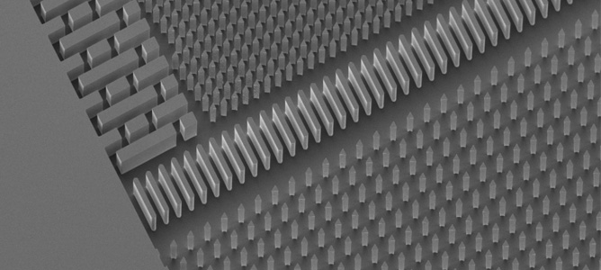
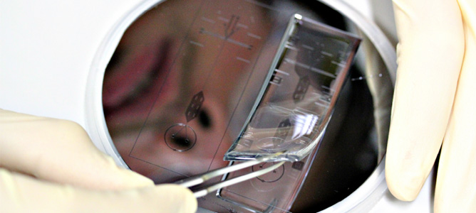
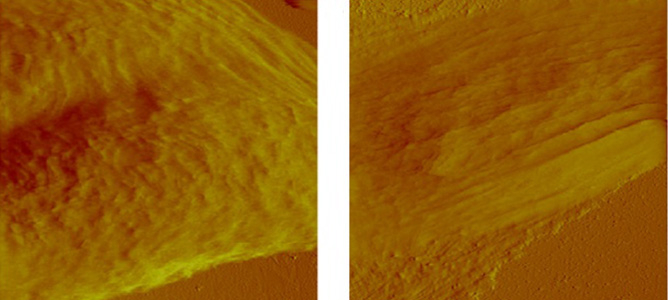
Nanostructural features present on a microfluidic wafer.
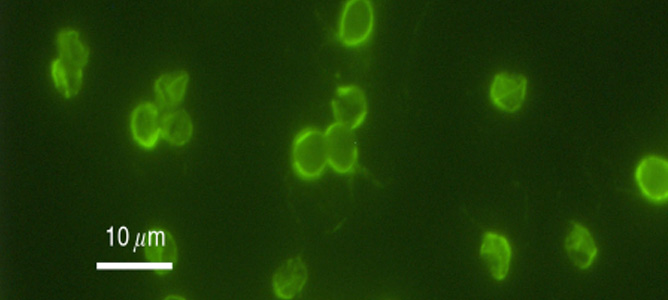
Cryptosporidium Parvum Bacterium
Close up of nanostructural features present on a microfluidic wafer.
Safer surgeries with microbots
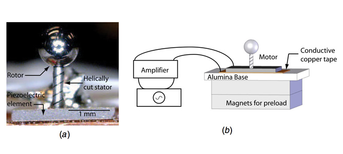
a) Photo of the micro-motor prototype showing the helically cut stainless steel ball as a rotor and the PZT element. b) Magnets were used to increase the friction coupling preload
Industry R&D looks at water source analysis
Automated systems for cell transfection
(a) Image of RTCM created using the Nanoprint MicroArray System located at MCN. (b) Close-up of Array Spots. Protein vectors are tagged with GFP and appear green. HeLa Cell Nuclei are labelled with DAPI and appear blue. DNA is cy3 labelled and appears red.
Synthesising high quality enzymes
Microfluidic device designs for the production of high quality enzymes
Identifying key materials for chemical sensors
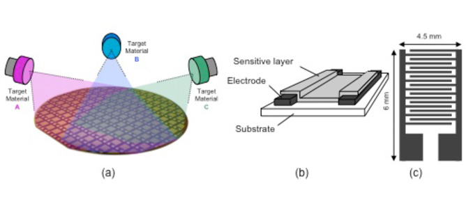
(a) Set-up for combinatorial sputter approach of metal oxide thin films, (b, c) cross- sectional view of the fabricated sensors.
50nm biosensors to detect antigens
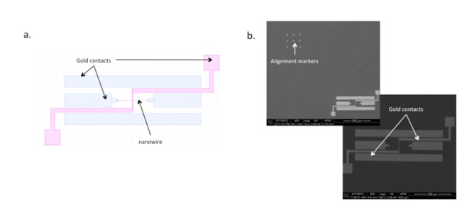
a) CAD file outlining the position of the alignment markers with respect to the single device; b) SEM images of the device with markers, and a close up on the device;
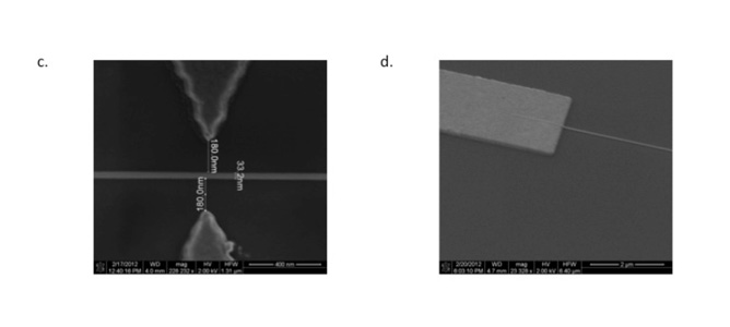
c) SEM image of the nanowire, less then 35nm wide, placed in between the electrodes very accurately. d) SEM image of the device dry-etched to the desired depth.
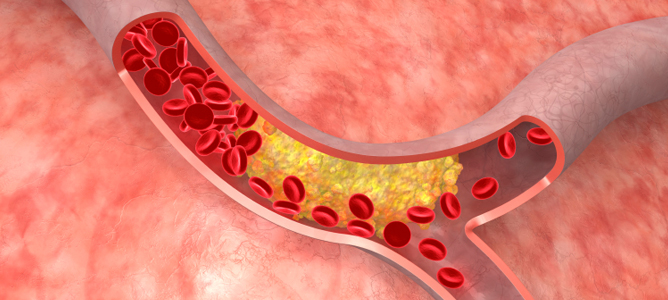
Predicting artery plaque rupture
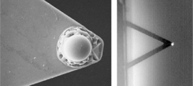
Sphere (left) attached to the cantilever tip (right).
AFM deflection image of cancerous (left) and benign breast cells (right).
Liquitab, not a hard pill to swallow
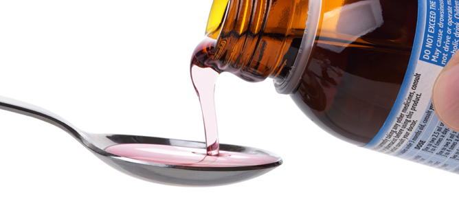

The liquitab will potentially assist millions in taking their daily medications.
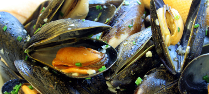
Enhancing Victorian mussel farms’ profitability
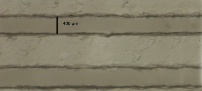
1: 3-dimensional polymer structure for research in mussel farming

