Detecting chemicals and biological contaminants
October 2011
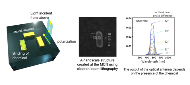
In his Fellowship project, Dr. Tim Davis from CSIRO is trying to develop new ways of detecting chemicals and biological contaminants, for applications in environmental monitoring and in biomedicince. As different chemicals interact with light in different ways, he is investigating methods of using light to detect and identify the different chemicals and biological substances.
The key issues are discrimination - being able to distinguish between similar chemicals or biologicals - and sensitivity - being able to sense low concentrations. The project aims to address this problem by controlling the properties of light at the nanoscale which Dr. Davis hopes will lead to better molecule discrimination and sensitivity. In particular, he will interact light with metallic nanostructures – these act like antennas for light.
The facilities at the MCN will allow the fabrication of the optical antennas at very small size scales, well below the wavelength of light, as well as the combination of them to control the light properties to study how it interacts with molecules.
This work will lead to new ways of detecting contaminants in the environment and new methods for screening for disease. Moreover, the technology has applications in other areas such optical communications and improving solar energy conversion.
Improving cancer therapies
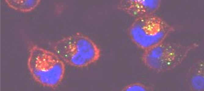
Confocal microscopy image showing the uptake of fluorescently-labelled pore forming proteins (green) by cancer cells (membrane in red, nuclei in blue).
October 2011
The ability to efficiently deliver large therapeutic molecules into cancer cells without harming healthy cells remains a largely unsolved technological challenge. Dr. Christina Cortez Jugo from Monash University aims to address this problem by developing sophisticated, nanoscale therapeutic carriers, with improved efficacy and specificity. Her Fellowship project will investigate the use of pore-forming proteins, which have the ability to temporarily puncture cells, in order to facilitate the transport of therapeutic molecules into cancer cells.
The scientific and technological advances in drug delivery and nanomedicine that would be enabled by this work are poised to revolutionise cancer treatment and lead to improved healthcare for many Victorians.
Environmental monitoring with nanobio membranes
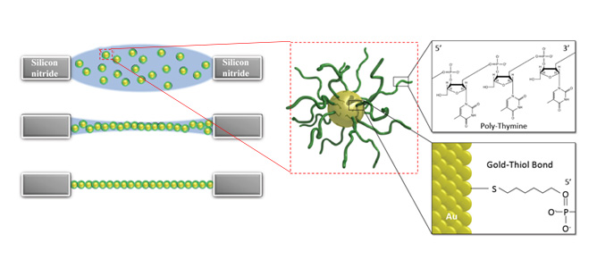
Drying of microconfined droplet of DNA-capped nanoparticles leads to the formation of the thinnest possible free-standing nanoparticle superlattice membranes.
October 2011
Associate Professor Wenlong Cheng’s Fellowship project aims to develop intelligent nanobio membranes which are as thin as possible for applications in environmental monitoring. He will address this problem by a combined top-down lithography (photolithography) and bottom-up self-assembly (DNA-programmable materials synthesis).
The photolithography, e-beam lithography and focused-ion-beam lithography tools as well as atomic force microscopy, scanning electron microsopy and confocal microscopy will be used in conjunction with the biological labs available at MCN.
It is hoped this work will lead to smart, ultrathin, multifunctional nanobio membranes and improved technologies in seawater desalination with low energy consumption, as well as at-home, lightweight, foldable toxic gas detectors with high sensitivity.
Bottom up methods for nano building blocks
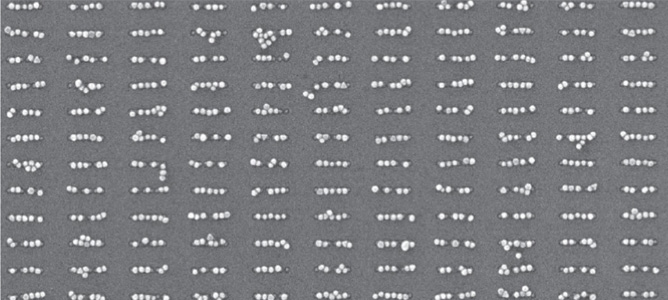
40nm gold nanoparticles, self-assembled onto surface–confined nanopatterns by means of DNA-directed self-assembly
October 2011
A lack of suitable bottom-up fabrication methods which allow for the assembly of custom-defined nanostructures impedes the integration of a plethora of new nanoparticulate building blocks into everyday devices in applications such as electronics, optoelectronics, sensing and photovoltaics. Associate Professor Udo Bach from Monash University is aiming to develop bottom-up fabrication methods for nanoparticles which have properties that are generally superior to lithographically defined structures.
He aims to achieve this by combining existing top-down nanofabrication techniques with novel self-assembly and printing methods to develop novel cost-effective ways of producing nanoparticle assemblies with well-defined configurations. To achieve this, he will utlise a series of MCN’s tools such as electron beam lithography, reactive ion etching and atomic layer deposition.
This work helps to build a knowledge base in a research area that is believed to hold the key for future manufacturing methods, while it is also useful for the improvement of novel photovoltaic technologies that are currently developed within the Victorian Consortium for Organic Solar Cells for the next generation of low-cost solar cells.
Evolving enzymes on a microchip
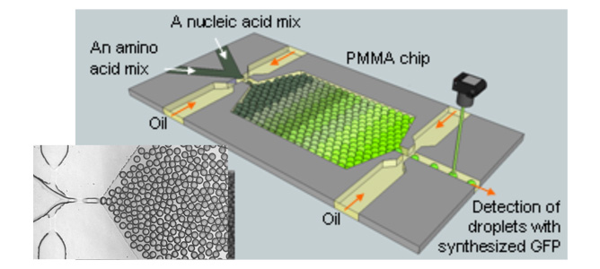
A picture of a microchip developed in CSIRO Microfluidics Lab for synthesis of a green fluorescence protein molecule
October 2011
Dr. Yonggang Zhu’s Fellowship project aims to develop an integrated microfluidic chip that can perform in vitro enzyme synthesis and evolution. This technology could potentially revolutionise nano- and bio-technology development.
The current protein evolution protocol requires screening of ‘libraries’ of massive numbers of candidate gene/enzyme variants, at a cost between millions and billions and take several years to complete. This project proposes to use microfluidic water-in-oil droplets to compartmentalise these libraries. These discrete micro-droplets are ideal bioreactors in which enzymes can be synthesised, screened and selected. The high level of controllability and speed of droplet production in microchips (~103 drops per second) allows for unprecedentedly fast and low-cost screening of massive libraries, with a potential 106-fold reduction in time and cost in comparison with existing technologies.
MCN’s state-of-the-art lithographical tools, characterisation tools and biological labs will be fully utilised for the project.
This work could potentially lead to a new way of developing novel biomolecules. Such a development would benefit many industry sectors such as biotechnology, chemical manufacturing, in vitro cell-free protein evolution, biomaterials, sensing and drug development.
Vertical arrays of gold nanorods achieved
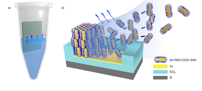
(a) Illustration of the experimental setup. The micropatterned substrate is placed in the centre of an Eppendorf tube and immersed in a concentrated colloidal solution of GNR’s. The substrate is left to dry under isothermal conditions. b) Schematic representation of the formation of standing arrays of GNR’s on a template upon solvent evaporation.
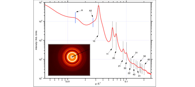
Transmission SAXS pattern recorded on a 100 μm × 250 μm section. Gray lines indicate the positions of the hcp reflections (labels are Miller indices (hk) for the reflections; not all are shown for clarity). The inset shows the associated diffraction pattern.
October 2011
Fabrication of novel self-assembly metallic nanostructures shows great potential for applications in biosensing, optical analysis, computing and solar energy conversion.
Working collaboratively with Instrument Manager Matteo Altissimo, Thibaut Thai of Monash University has combined bottom-up self-assembly processes with top-down techniques to generate vertical arrays of gold nanorods on patterned substrates.
Gold nanorods (GNRs) are of particular interest to researchers as they display unique yet highly variable optical properties. One such property is their remarkable sensing ability, which is exploited during Surface Enhancement Raman Spectroscopy (SERS). During this process GNR’s are closely packed together in a hexaganol-type arrangement forming high density “hot- spots”. If a molecule is within this “hot-spot”, its Raman signal will be greatly amplified.
The Raman signal then acts as a molecular fingerprint and is key in characterising a materials’ chemical structure, temperature or frequency mode.
In past research, controlling the self-assembly process has been a challenge due to the anisotropic nature of the nanorods.
Using a method developed at MCN, researchers were able to control the orientation of the assembly and select a precise location to express the desired features. The mask aligner, reactive ion etching process, electron-beam evaporator and scanning electron microscope were all critical components during the fabrication process.
The accuracy of the micro-patterns was confirmed using small-angle X-ray scattering (SAXS), a characterisation process undertaken using the SAXS beamline at the Australian Synchrotron National Research Facility.
In addition to showcasing the vast array of fabrication and characterisation tools available within MCN, the work highlights the unique relationship between the Australian Synchrotron and MCN, acting in unison as a hub for world-class research.
Cantilevers to assist with fluid characterisation
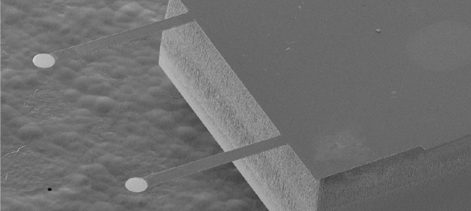
Scanning Electron Microscope Image of Si Cantilever
October 2011
MCN’s Doug Mair has worked collaboratively with Dr Raymond Dagastine from the Particulate Fluids Processing Centre at Melbourne University to develop silicon cantilevers to continue the already extensive research performed by his group into the area of bubble and oil drop dynamics. The goal of the project was to design and manufacture silicon cantilevers with different spring constants and gold paddle sizes to assist with characterisation of deformable drops on Confocal and AFM systems.
The project went through several stages, from concept to design through to process development. All process development has been completed and the first wafer of cantilevers has been successfully fabricated and tested. This project brought together many of the capabilities of MCN including front side and backside pattern alignment, chrome/gold evaporation, deep reactive ion etching (Bosch Process), chemical processing through to final characterisation of product on AFM all under the one roof.
The project has been a great success and opens the door to a large range of cantilever based designs.
Manipulating hot spots to increase biosensor sensitivity
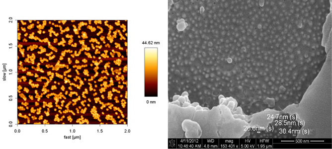
(a) A 2μm2 AFM image of gold nanoparticles on a flat substrate. (b) SEM micrograph showing measurements of nanoparticles under a polymer film.
October 2011
Surface Plasmon Resonance (SPR) can be described as the resonant, collective movement of electrons activated by incident light that travels in a direction parallel to a surface. When two nanoparticles are placed in close proximity, the electric field is greatly enhanced creating a ‘hot spot.’ These hot spots are of particular interest to researcher Soon Ng, of Monash University. Working collaboratively with MCN’s Varsha Lal and Matteo Altissimo, Soon is manipulating these electric field hot spots to increase the sensitivity of his biosensors.
The JPK Nanowizard II AFM at the MCN was used to accurately measure the height of the particles as well as the overall topography. One of the key features of this instrument is that it allows imaging in both air and liquid.
Additionally, the FEG-SEM was used to measure the size of particles and to aid in the calculation of surface coverage. Future work on this project will utilise electron beam lithography to create templates for nanoparticle absorption in order to study the spatial configurations related to the hot spots. As well as having applications in biosensing, chemical detection and solar cell technologies, the properties of surface plasmon resonance may be used during characterisation and imaging processes.
This is an example of a project where multiple capabilities available at MCN were used in sequence and assembly and characterisation.
Photonic circuitry from the noble metals

FIB fabrication of linear arrays of silver nanocrystals.

FIB fabrication of linear arrays of silver nanocrystals. Scale bar for the top and middle images are 1000nm and scale bar for bottom image is 100nm.
October 2011
This project looks at fabricating linear arrays of nanocrystals by using the FEI Helios NanoLab 600 Dual-Beam Focused Ion Beam-Scanning Electron Microscope (FIB-SEM) to test the capability of the arrays to act as nanoscale optical fibres. MCN’s Dr. Manoj Sridhar, collaborated with Dr. Alison Funston from Monash University to compare this capability with existing nanowire technology.
One of the major challenges for the use of light-based circuits remains the confinement of the physical extent of the light to sub-wavelength dimensions without compromising the distance the light energy can travel.
Together, they were able to fabricate linear arrays of silver nanocrystals using a combination of chemical synthesis and nanofabrication. Tests are currently underway to assess the waveguiding capability of the structures. Initial results show structures created by FIB are able to act as waveguides. The capabilities at the MCN were essential to this project to fabricate the arrays by exploiting the small diameter of the FIB-SEM instrument.


