Micro sensors to monitor blood pressure

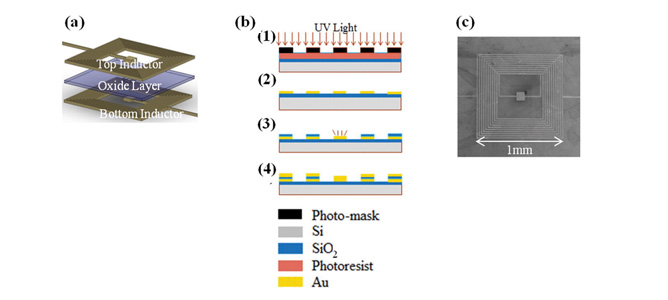
 (a) An exploded view of the double layer planar micro inductors, (b) A summary of the fabrication steps: (1) Oxide deposition and UV Lithography, (2) E-beam evaporation and lift-off. (3) SiOdeposition and etching. (4) Steps 1–2 repeated for the second inductor layer, (c) A scanning electron micrograph of the resulting coil. 

October 2013

Hypertension, if left untreated, can lead to a number of health problems including strokes, heart failure and kidney failure. Regular monitoring of blood pressure is currently inefficient and is not performed as regularly as necessary to monitor and mitigate these serious health concerns.

Led by Dr. Tuncay Alan, researchers from Monash University are developing a highly sensitive, implantable blood pressure sensor. These micro-sensors could be implanted into patients suffering high blood pressure to better monitor levels. This would allow continuous and routine blood pressure measurement and readings would then be communicated wirelessly to a hip-held alert unit. The results could then lead to earlier diagnoses of related diseases, complications and more efficient treatment.

The devices being investigated by Dr Tuncay Alan and  Associate Professor Neild in the Laboratory for Microsystems, are inductor sensors which are far more sensitive when downscaled than other sensors which have previously been researched. The team have developed a model which uses two planar spirals separated by an insulating layer, and have fabricated double layered microplanar coils at MCN. The opposing magnetic fields between pairs of these coils create a high sensitivity to relative displacement which can be linked to pressure. Results so far show that device dimensions could be shrunk for further improvements on resolution. Overall results indicate that inductive sensing has a good potential for micro scale sensing applications. Collaborators are currently investigating the circuitry necessary for the sensor and the fabrication of a complete system.

You can read more about this project in Nanoscale displacement sensing using microfabricated variable inductance planar coils, published in Applied Physics Letters.

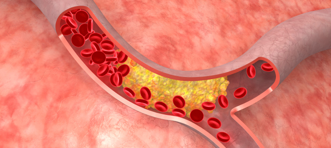
Predicting artery plaque rupture

Lipid build up in artery walls can cause plaques to form that can rupture and cause significant health problems.

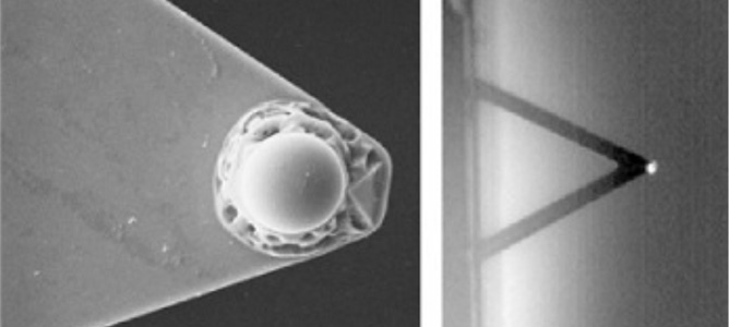
Sphere (left) attached to the cantilever tip (right).

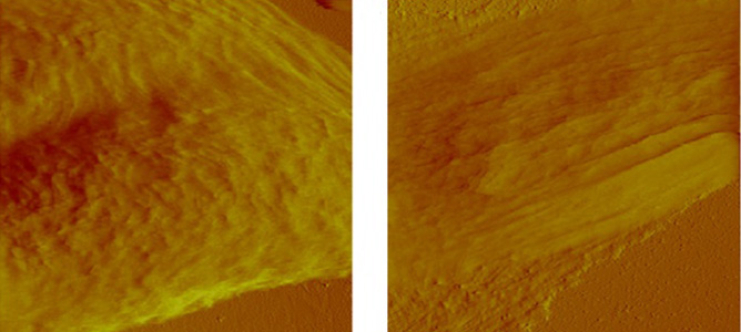
AFM deflection image of cancerous (left) and benign breast cells (right). 

February 2012

Due to lipid build up in artery walls, plaques form that can rupture and cause significant health problems for patients. Researchers at the Baker institute have worked together with MCN to study and predict the likelihood of plaque formation and rupture due to Atherosclerosis in an attempt to prevent it.

Atherosclerosis occurs when lipid steaks infiltrate and build-up in the artery walls, creating a plaque as a result. These plaques can be divided in two distinct types: those that rupture (vulnerable) and those that are less likely to rupture (stable).

In this project, the Nanowizard II AFM was used to determine the elastic properties of the vessels and plaque as well as the topography of the plaque surface. The first results were directly implemented in computational fluid dynamics software. Studying the surface topography then led to the development of models for the boundary conditions along the artery walls. This work will employ a new model in mice, to be used for predicting the formation and properties of vulnerable plaques.

A difficulty in the present approach is working with wet and soft samples. In particular, working with soft samples implies a specific set up for modified cantilevers, in which a sphere is bound to the AFM cantilever (see image above). Tests will be conducted using breast cancer cells to improve this methodology and optimise the conditions for cantilevers. Cantilever optimisation experiments will utilise the cleanroom facility at MCN.

Cantilever-based biosensors to help detect cancer antibodies

The biosensor could detect cancer biomarker antibodies to assist with developing cures for cancer 

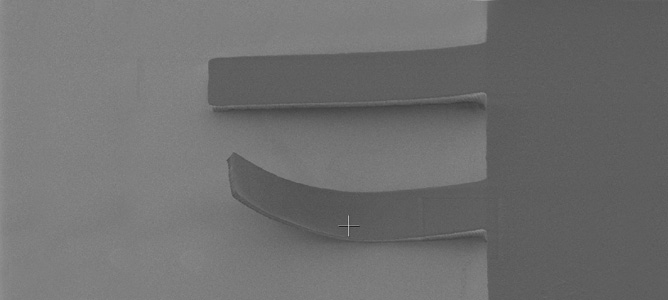
SU8 Cantilever – second arm shows signs of thermal stress.

October 2011

Utilising a photoactive material, researchers at MCN and Deakin University are designing a cantilever based biosensor which detects cancer biomarker antibodies. The cantilevers are to be made from a photoactive material called SU8. MCN houses all the required tools under one roof that allows manufacture and testing of the complete cantilever device. Development so far has concentrated on two steps, photolithography and a special co-polymer layer.

The cantilever is created in two stages, a thin, 2µm  layer which forms the cantilever arm, and a thick, 100µm  layer which acts as the cantilever body, which are manufactured on silicon wafers. SU8 bonds to silicon so a co-polymer layer must be deposited onto the silicon surface in the MCNs reactive ion etchers, which forms a weak layer that allows the structure to be extracted from the silicon surface.

Development of these two stages is ongoing. After this the cantilevers will require a gold layer on the back to form a reflective surface required for subsequent characterisation steps. Prior to characterisation the devices will be taken to Deakin to have the surface functionalised to allow detection of cancer biomarker antibodies. Following this step the product will be returned to MCN to perform characterisation and verification within the biological atomic force microscope.

Understanding emulsions and foams

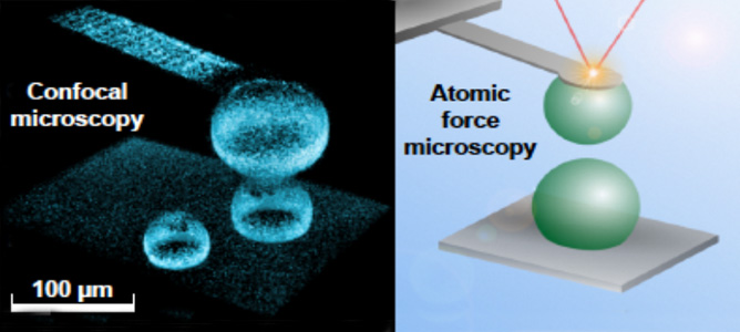
Confocal image of two oil drops immobilized in an atomic force microscope. (top right) Schematic of the measurement where a custom micro-fabricated cantilever is used to hold the top drop in position. 

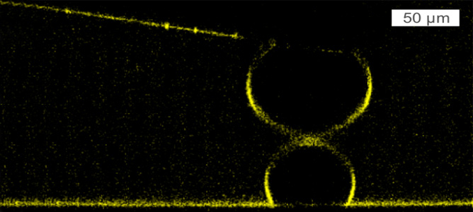
Vertical slices of a confocal microscopy image showing the profile of the two drops when separated far apart and when deformed.

October 2011

This project, led by Professor Ray Dagastine from the University of Melbourne, looks at fabricating nano- and micro-sizes devices to integrate with atomic force microscopy with the aim of developing new tools to study the complexity of interfaces in soft materials. The collisions between drops or bubbles in soft materials such as emulsions and foams control macroscopic behavior in growing sectors of biotechnology and nanotechnology as well as in materials used in everyday items. Some examples include the production of salad dressing, milk and shampoo, industrial processes such as solvent extraction used for pharmaceutical and mineral purification, as well as development of micro-fluidic devices for new applications.

The fabrication of custom designed micro-cantilevers allows the manipulation of oil drops or bubbles during these collisions to better understand the fundamental behaviors in emulsions and foams. Currently, these types of devices can only be fabricated internationally. In combination with the device fabrication, the atomic force microscope has been integrated with a laser scanning confocal microscopy to give real-time images of the collisions between drops and bubbles in high resolution. The nanoscale forces between these soft objects are sensitive to the addition of surfactants, polymers and biological molecules at these interfaces. This imparts a multilevel complexity and lateral heterogeneity starting at the molecular scale and spanning to the macroscopic scale. This complexity often creates a gap in our understanding of how these molecular structures mediate dynamic interfacial forces on the nanoscale and the material behavior on a large scale.

Cantilevers to assist with fluid characterisation

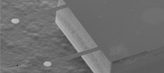
Scanning Electron Microscope Image of Si Cantilever

October 2011

MCN’s Doug Mair has worked collaboratively with Dr Raymond Dagastine from the Particulate Fluids Processing Centre at Melbourne University to develop silicon cantilevers to continue the already extensive research performed by his group into the area of bubble and oil drop dynamics. The goal of the project was to design and manufacture silicon cantilevers with different spring constants and gold paddle sizes to assist with characterisation of deformable drops on Confocal and AFM systems.

The project went through several stages, from concept to design through to process development. All process development has been completed and the first wafer of cantilevers has been successfully fabricated and tested. This project brought together many of the capabilities of MCN including front side and backside pattern alignment, chrome/gold evaporation, deep reactive ion etching (Bosch Process), chemical processing through to final characterisation of product on AFM all under the one roof.

The project has been a great success and opens the door to a large range of cantilever based designs.