Next generation pathogen detection with lab-on-a-chip platforms
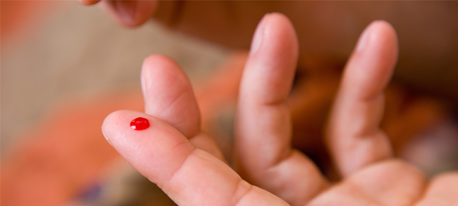
Taking a sample could be as simple as a fingerprick of blood, as is common in blood sugar monitoring.
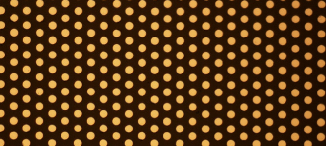
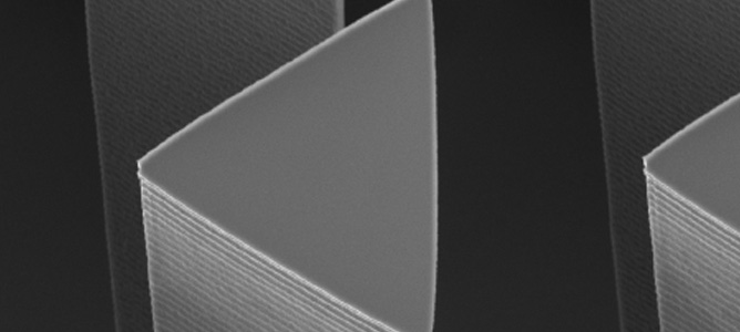
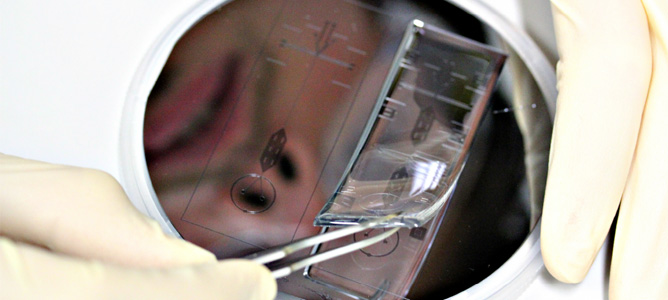
The metal microdiscs sputtered on a glass slide at MCN. The posts are 5 µm in diameter and 300 nm high.
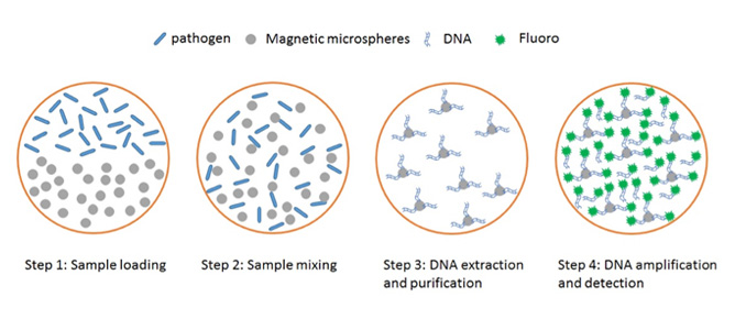
The schematic drawing of the pathogen detection: DNA extraction, purification, amplification and detection.
Microchips to mimic living cells
A microchip that can generate microdroplets for protein and nanoparticle synthesis.
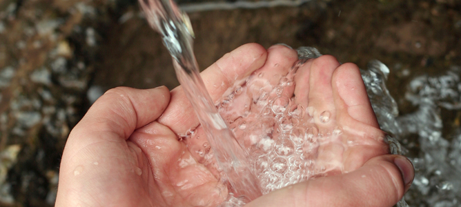
Protecting the nations’ water supply
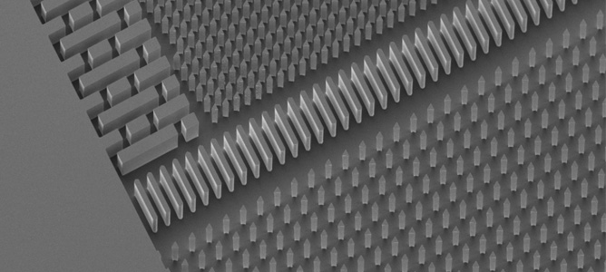
Nanostructural features present on a microfluidic wafer.
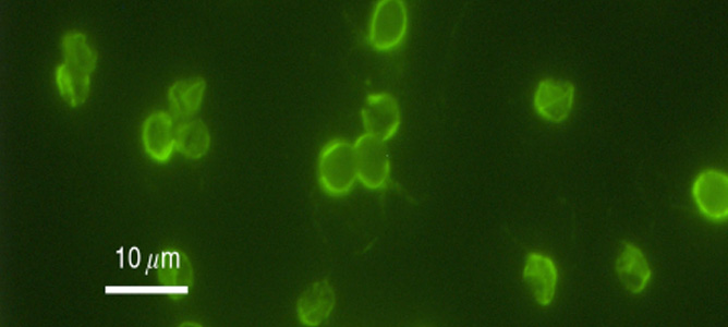
Cryptosporidium Parvum Bacterium
Close up of nanostructural features present on a microfluidic wafer.
Synthesising high quality enzymes
Microfluidic device designs for the production of high quality enzymes
Acoustic nanofluidics
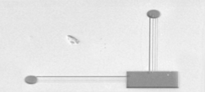
Fluid reservoirs are connected with an output well via 100, 50, and 20nm wide channels in lithium niobate. Upon filling the reservoirs and starting acoustic radiation in the substrate, the channels rapidly fill the output well at speeds many orders beyond physically predicted rates. This entire structure would fit in this full stop.
Evolving enzymes on a microchip
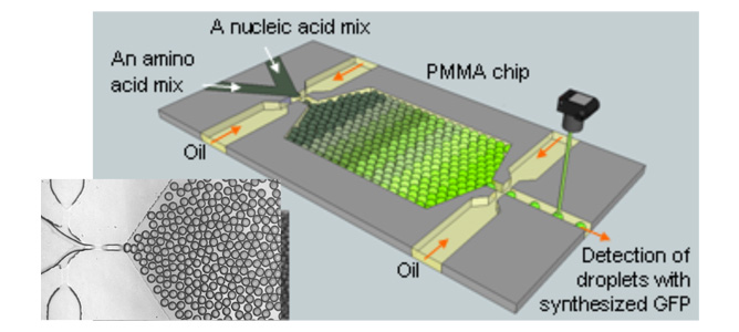
A picture of a microchip developed in CSIRO Microfluidics Lab for synthesis of a green fluorescence protein molecule

