Nanofibre facemasks provide a breath of fresh air

Deakin University’s Institute of Frontier Materials (IFM) has been working with Xinau Technology to develop a new type of nanofibre facemask that will replace traditional microfibre-based equivalents.
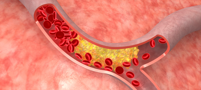
Air quality is on the decline in huge areas of the developed and developing world. The International Energy Agency attribute an estimated 6.5 million deaths per year to indoor and outdoor air pollution – it is reportedly the fourth largest threat to human health, after high blood pressure, dietary risks, and smoking. Pollutants that measure less than 2.5 microns (PM2.5) are a particular concern, their small size means they are able to penetrate deep into the lungs.
Facemasks are now commonly used in heavily affected areas, but conventional microfibre masks either don’t provide the necessary filtration or block air ow too heavily, making it hard to breathe.
As “smog seasons” become a regular occurrence, cities such as Beijing are seeing a huge number of residents turn to expensive, high-end masks to help them breathe easy.
A team at the IFM are now using ANFF VIC’s electrospinning equipment to create nanofibre-based masks that are drastically better at stopping inhalation of PM2.5 particles and provide very little breath resistance. The pilot electrospinning machine is able to prepare 2-metre-wide nanofibre sheets in a continuous manner and has a production capability of up to 1,000m2 per day.
“ANFF VIC’s needleless electrospinning machines at Deakin university made it possible to process large-size nanofibre nonwoven sheets with consistent structure.” – Professor Tong Lin
“When a layer of nanofibre nonwovens is inserted into conventional facemask, the breath resistance did not increase much, however, the capture efficiency for PM2.5 was significantly improved,” Professor Tong Lin, leader of the team at Deakin University, said. This means the team can make the filtering material thinner and more breathable, while still stopping up to 95% of airborne pollutants.
The key component to maintain high PM2.5 filtration efficiency is the nanofibre sheets, which are highly porous and have a large surface area.
“ANFF VIC’s needleless electrospinning machines at Deakin university made it possible to process large-size nanofibre nonwoven sheets with consistent structure,” Tong continued. “It also allows you to adjust the fibrous structure by changing the operating parameters such as voltage, spinning distance and polymer components.”
The team are currently working with Xinau to commercialise the technology.
Exploring gold nanowires and nanorods
Professor Wenlong Cheng from Monash University has been exploring unique properties of gold nanomaterials for over 16 years. Two recent projects, conducted at ANFF-Vic’s Melbourne Centre for Nanofabrication (MCN), involved gold nanorods and gold nanowires.
Wearable sensors
Stretchable, conductive materials are driving the capabilities of tomorrow’s soft and wearable technologies.
Wearables may hold the key to accurate and comfortable health monitoring and movement detection. There’s also the potential for a range of intuitive human/machine interfaces to be created that could ultimately enable implantable biomedical devices.
Wenlong and a team of researchers from Monash University have been making a range of flexible sensors by coating elastic materials with interwoven networks of conductive ultra-thin gold nanowires (UGNWs). According to Wenlong, the UGNWs are the world’s thinnest gold nanowires at only 2nm wide — a DNA double helix, by comparison, is just over 2nm in width.
The elastic substrates that the UGNWs are grafted onto are flexible and biologically safe, making them ideal candidates for wearable electronics.
Researchers worked at MCN to create wearable pressure sensors, skin-attachable strain sensors, flexible transparent electrodes and stretchable supercapacitors.
To create a sensor, UGNWs are grown via a solvent-based chemical process that yields a host of hair- like ultra-thin fibres. Once in a liquid suspension, the wires are “painted” onto the elastic material, forming
an interwoven mesh of electrically conductive fibres once the paint dries. When the UGNW-coated elastic
is stretched, the device’s electrical resistance changes too, providing
a real-time and reversible electrical representation of the sensor’s state.
Wenlong exploits these changing properties to detect and monitor movement with an incredibly high degree of precision. His devices offer touch sensitivity and can pick up the tiny forces associated with artery wrist pulses.
“They are able to resolve pressing, stretching, bending, and torsion
forces as well as acoustic vibrations,” Wenlong said. The team’s devices are cheap to manufacture and attachable to almost any surface, allowing accurate measuring of skin or muscle deformation anytime, anywhere.
In the health sector, for example, increased sensitivity of these devices could reveal previously undetected heart defects or be used to monitor detailed hand movements in robotic infrastructure such as advanced prosthetics.
Targeting tumours
At present, the vast majority of cancer treatments work by attacking cancer cells but often affect healthy tissue in the process. The approach, whilst effective, produces a large number of side effects including irreparable and extensive tissue damage, hair loss and nausea, among others.
There has therefore been a great deal of effort devoted to improving the selectivity of cancer therapies in order to minimise collateral damage. Increasingly, the idea is to hunt with a sniper rifle, rather than a shotgun.
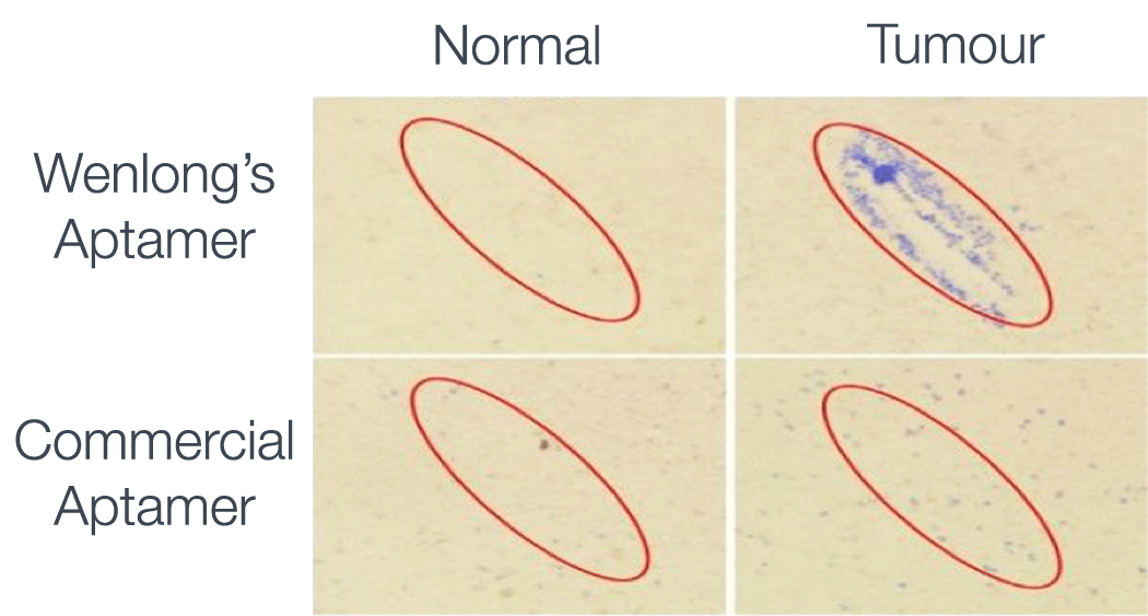
Wenlong and his colleagues have gone a step further and developed what is effectively a guided missile that exclusively binds to target tumours before heating defective tissue to the point of destruction. Advancing a treatment called photothermal therapy, the method uses gold nanorods (GNRs) – shorter, wider versions of gold nanowires – which rapidly heat up to 45°C when irradiated by near-infrared (NIR) light.
To specifically target the cancerous cells, the GNRs are coated with DNA-specific aptamers that Wenlong selected to identify DNA unique to
the tumour cells. Aptamers are single strands of DNA that bind selectively
to a complementary piece of genetic material. The theory is that once the aptamer binds to the target tumour cell, the GNR is heated with the NIR light and the cell is destroyed.
Wenlong and his team collaborated with a team led by Professor David Jans, NMHRC Senior Principal Research Fellow at Monash University to test the GNR-aptamer “missile” on breast ductal carcinoma cells residing in healthy tissue.
Wenlong’s team was able to observe the selectiveness and efficacy of this process using the MCN’s hyperspectral imaging capabilities. The initial results indicate that once the GNRs had been heated by NIR light, 96 per cent of tumour cells had been successfully targeted and 71 per cent of them were destroyed, with less than one per cent of healthy tissue affected.
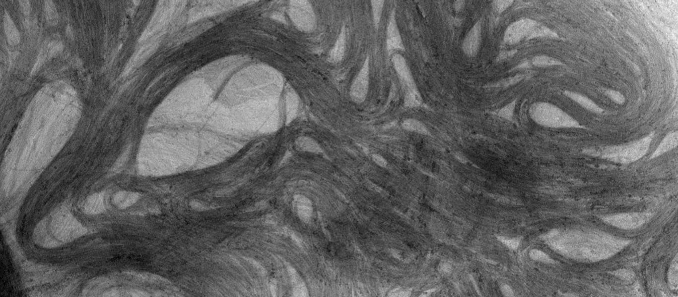
Folding plasmene nanosheets
Professor Wenlong Cheng has been investigating how to make an unusual class of materials with exotic properties and unprecedented real-world applications.
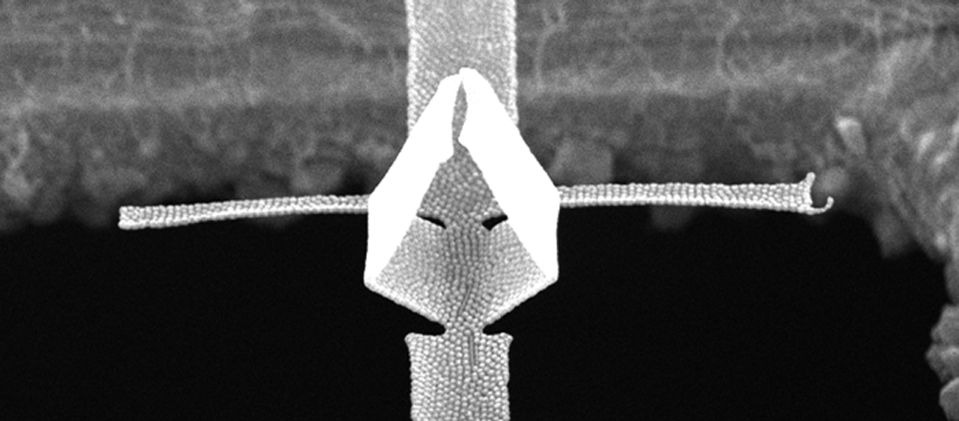
Formally introducing plasmene nanosheets to the world, he and his team have created self-folding sheets of nanoparticles that can change the behaviour of light.
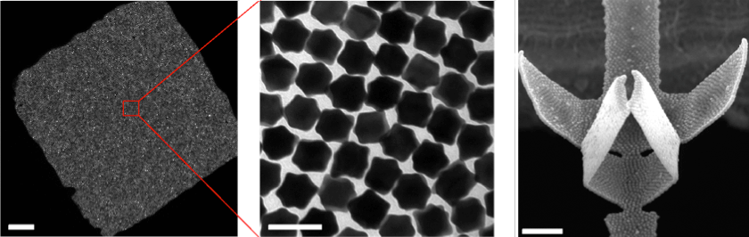
The nanosheets consist of a superlattice of nanoparticles, organised in a hexagonal pattern much like the arrangement of carbon atoms in the wonder-material, graphene.
Wenlong’s team creates the materials by sparsely distributing nanoparticles covered with protruding polystyrene hairs, or ligands, across the surface of a water droplet. By evaporating the water the surface area shrinks, bringing the particles closer together. Attractive van der Waals forces between the nanoparticles draw them together while repulsive forces between the ligands manoeuvre the nanoparticles into the desired hexagonal configuration.
The nanoparticle-laden water droplet is suspended over an open cavity, in this case a hole in a copper sheet, which gives the nanosheet a place to form. Once all the water has been removed, a continuous nanosheet forms neatly across this cavity like the membrane on a drum, allowing Wenlong to begin to play with it.
Using ANFF-Vic’s focused ion beam (FIB) instrument at the Melbourne Centre for Nanofabrication, the team “scores” specific patterns into the sheets, deliberately weakening them at key points. The weakened regions are akin to the paper folding lines introduced in origami, where the layout and severity of the folds define the final three-dimensional structures.
This scoring process means the resultant shape’s properties can be tuned by manipulating the pattern cut into them and the order in which the folding is performed, enabling tailoring of the final product to meet specific end applications.
Researchers have investigated the properties of this self-folding before, but the designs of the well-defined origami structures of this kind have not been realised until now.
Light interacts and reflects off differing origami shapes in a way that is unique to each contour. By changing the structure, Wenlong can exploit his plasmene sheets to perform some useful tricks.
One useful application for these tiny sculptures is in nanophotonics as plasmonic waveguides for directing and switching light at the nanoscale by circumventing diffraction limits.
The plasmene nanosheets produced can also be used as dual-coded encryption security labels as it’s almost impossible to fraudulently create the shapes without the initial blueprint. This poses enormous potential benefits to the banknote industry, which is always searching for more secure methods of deterring counterfeiters.
The plasmene sheet’s semitransparency, mechanical softness and uniform response to light suggest that it could be an ideal material in the production of secure identifying labels for chemicals, currency and commodities, among others.