Nanofabulous Seminar: A new imaging modality in nanomaterials and life sciences
Engineered or complex particles found in nano-electronics, photonic materials, and nano-sensors, etc. have experienced a rapid growth in research interest and application over the past two decades. Similarly, the interface between nanomaterials and biology has also grown rapidly. These outcomes are in part due to improving techniques to fabricate materials and biomaterials, from both top-down and bottom-up approaches, even at scale.
The characterisation needs for such materials leans heavily on existing microscopy or metrology tools that often balance resolution against the time, expense and complexity of measurement workflows. In addition, as objects get small, the methods become more specialised.
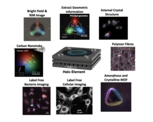
Motivated by the need to visualize complex particles and objects in engineered materials and dynamic processes in life sciences, this talk will focus on the operating principles of Resonance Imaging Microscopy (RIM). RIM is a new imaging modality to provide simple and easy characterisation of objects with nearly any shape and composition, ranging in size from hundreds of microns to tens of nanometres. RIM is a multi-source evanescent field scattering technique that enables high-speed, non-destructive, label and stain-free imaging in real time (up to hundreds of FPS) using visible light.
For particle metrology applications, we will present both direct and statistical results validating the fidelity of geometric measurements for a selection of spherical calibration particles with radii spanning four orders of magnitude. We will showcase more complex materials including nanorods and materials with complex internal structures. In life sciences we will show results of multi-modal characterisation studies that demonstrate how RIM is able to elucidate the internal structure of, and relationship between, untagged cells and bacteria. We will showcase how RIM can be used to visualise a range of other biological materials, both static and dynamic in nature. All achieved without the need of fluorescent proteins, dyes or conjugated antibodies or risks from photobleaching when examined using traditional laser-based microscopy systems.
Prof Raymond R. Dagastine
Department of Chemical Engineering, The University of Melbourne, VIC 3010, Australia
Tiny Bright Things, Carlton Victoria 3053, Australia
11:00am, 15/08/2024
Melbourne Centre for Nanofabrication
151 Wellington Road, Clayton, 3168
Zoom link: click here
Meeting ID: 858 5837 9980 and passcode:873578

