Microscopic Gardening wins Image of the Year 2018
It appears that the ANFF-VIC community has green fingers this year – Microscopic Gardening: Tiny Blossoms of Silicon by Vivek Garg has been voted the ANFF-VIC Image of the Year 2018.
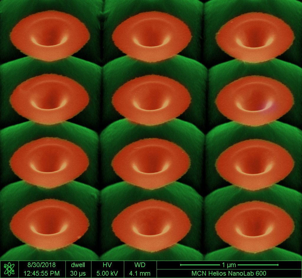
The image shows a scanning electron micrograph (false-color) of Silicon (Si) nanoflowers, created using MCN’s Focused Ion Beam (FIB) in conjunction with wet chemical etching.
As winner of the competition, Vivek will take home a $200 prize.
Vivek and his colleagues are investigating fabrication of 3D freeform structures of Si, such as these nanoflowers, due to their unique optical properties. Such structures can be engineered to selectively absorb light, and produce various colours depending on their architecture – they have tremendous potential for future optics applications such as optical security, polarimetry, and spectral imaging.
“The bulk structuration of Si substrate, based on the ion implantation design and area, allows fabrication of exotic functional and 3D micro/nanostructures on Si substrate exhibiting unique optical properties for applications in nanophotonics and physical sciences,” Vivek explained.
Vivek is a PhD candidate with the IITB-Monash Research Academy, a collaboration between IIT Bombay, India and Monash University, Australia. He is working with Dr Rakesh Mote (IIT Bombay) and Dr Jing Fu (Monash) on the fabrication and controlled manipulation of freeform 3D micro/nanostructures with ion beams. This work is a part of his thesis project, in which he is investigating the use of FIB nanofabrication in creating novel nanostructures for diverse applications such as anti-reflection, colour filtering, sensors and more.
Read more about Vivek’s work here http://www.vivekgarg.org/, or view the full shortlist for the 2018 Image of the Year competition below.

The 2018 ANFF-VIC Image of the Year
Here is the shortlist for the ANFF-VIC Image of the Year, click on the images to see the original.

“Aboriginal art”
(Confocal fluorescence microscope) – Nazia Tabassum
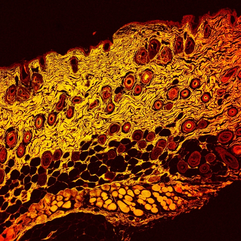
'Mice skin histology: Confocal fluorescence microscope image showing different viable strata; top stratum corneum, epidermis, dermis and hypodermis, containing different cells such as sebaceous glands, hair follicle bulbs, adipocytes cells, panniculus carnosus and pigmentation in bulbs. This histology was conducted after topical delivery of fluorescent molecules using silicon nanoneedles. In future studies, this designed study will facilitate to investigate the penetration and permeation of fluorescent-labelled antineoplastic drug into viable layers of skin and drug delivery against skin cancer melanoma.'
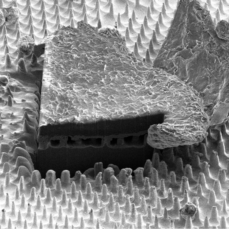
"Rivalry"
(SEM) – Aaqil Rifai, Alastair Stacey, Kate Fox
'Polycrystalline diamond (PCD) coated on 3D printed titanium using the MCN CVD 6500 equipment. This is an SEM image which beautifully represents the 111 (triangular) facets training whereas the 100 (square) facets do not. Our work has been published in the article entitled: "Polycrystalline Diamond Coating of Additively Manufactured Titanium for Biomedical Applications".'
"NanoPiano"
(SEM) – Marina Adeeb Tadros
'It is Fibroblast cells plated on silicon nanowire. The image was taken at MCN at FEG-SEM.'
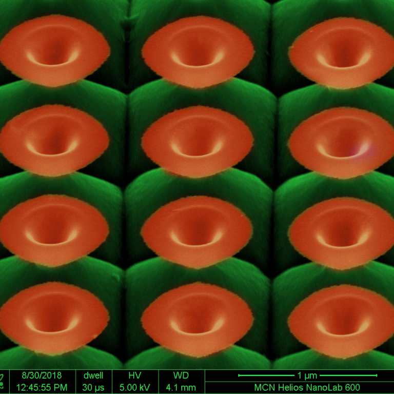
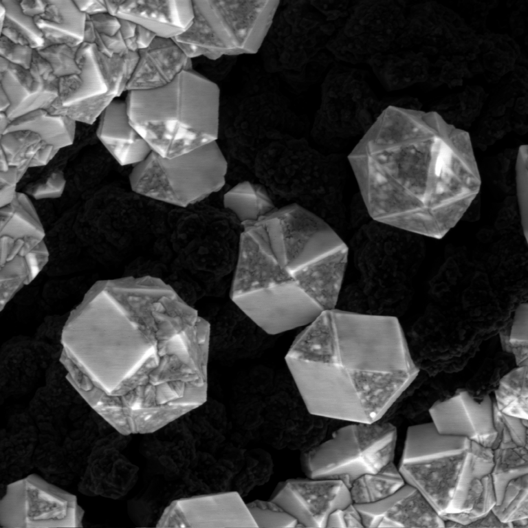
“Microscopic Gardening: Tiny Blossoms of Silicon” – Winner
(SEM) – Vivek Garg
'The image show scanning electron micrograph of Silicon nanoflowers realized with focused ion beams in conjunction with wet chemical etching. The bulk structuration of Si substrate, based on the ion implantation design and area, allows fabrication of exotic functional and 3D micro/nanostructures on Si substrate exhibiting unique optical properties for applications in nanophotonics and physical sciences.'
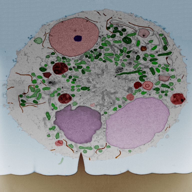
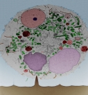
"The Tree of Life"
(SEM) – Stella Aslanoglou, Crystal Chen, Gediminas Gervinskas
'Scanning electron microscopy (SEM) image of a focused ion beam cut revealing the interface between a mouse immune B cell and a single silicon nanowire after 1 h of incubation. Silicon nanowires (SiNWs) are employed for the intracellular delivery of biomolecules towards the development of a novel cell-based immunotherapy. SiNW fabrication and cell culture was performed at MCN facilities. FIB-SEM sample preparation and imaging was performed at the Ramaciotti Centre for Cryo-Electron Microscopy.'